Research
- Copenhagen Center for Biomedical Quantum Sensing (CBQS)
- Fundamental Physics I: Short-Time Brownian Motion
- Fundamental Physics II: New Tests of Quantum Mechanics
Guiding Principles and Philosophy
The Raizen lab follows two parallel paths: the pursuit of discovery of fundamental physics and the development of new applications that bring benefit to humanity. The paths are intertwined; one enables the other. Our research is conducted by a highly motivated team working on tabletop experiments.
The Case for Tabletop Physics (with thanks to Jason Boynewicz)
Remarkably, a small group of people can change our entire understanding of the universe, with an experiment that fits on a single table. This is the great historical legacy of the field, from the Michelson-Morley experiment to all the atomic physics experiments that defined our understanding of quantum mechanics.Our lab adheres to the romantic vision of tabletop physics, a true call back to the days of breakthroughs by small teams. From testing quantum mechanics to understanding the microscopic nature of fluids, our tabletop experiments are pushing the boundaries of knowledge.
Meeting Challenges from the Past
The rich history of physics discovery is punctuated by singular scientists of great stature. Maxwell, Einstein, Rutherford, Weinberg, and many others, all accomplished great things in their lifetimes. They also left us with challenges as guideposts from the past. Maxwell wanted his thought experiment, Maxwell's Demon (1871), to be realized. Einstein proposed to probe Brownian motion on ultra-short time scales (1907). Rutherford wanted to find an alternative to radiochemistry (1911). Weinberg envisioned new physics beyond quantum mechanics. Our group is inspired and motivated to meet these challenges with our current and pending experimental breakthroughs. We are constantly searching for other historical challenges to meet, and to pose new challenges for future generations of scientists.
Physics to Benefit Humanity
The Novo Nordisk Foundation in Denmark shares and supports our vision; we have a responsibility to use advances in physics to benefit humanity. One direction is the development of new and efficient methods to produce stable and radioactive isotopes of the elements. There are important applications in diagnosis and treatment of metabolic disorders, cancer therapy, and early diagnosis of disease.We are also developing ultra-sensitive methods for acoustic detection near the quantum limit of sensitivity for medical imaging, and ultra-sensitive detection of molecules using recent advances in nanotechnology. This work will move from the research lab to The Pointsman Foundation, www.pointsman.org, a non-profit foundation which will implement inventions for the world.
Current Research
Copenhagen Center for Biomedical Quantum Sensing (CBQS)
Prevention of Iron Deficiency
Iron is an essential mineral for our health, needed for transporting oxygen to the brain and the body. Iron deficiency and the resulting anemia are widespread, affecting around two billion people worldwide, with a very high rate in children and in women during pregnancy due to the increased iron needs of these populations. It is considered a crisis in pediatric healthcare, due to the dire effect of iron deficiency on 50% of children worldwide and 15% of children in the US alone. Iron deficiency can cause severe developmental problems and permanent brain damage to infants and toddlers and decrease school performance in older children.
Although abundant in nature, iron is difficult to absorb from food, especially for young children where the need for the element is most critical and its lack potentially devastating and permanent. Even in affluent countries like the US, this problem is frequently undiagnosed and difficult to treat effectively.
Our collaborator and pediatrician Dr. Steven Abrams pioneered the use of completely safe, naturally occurring rare isotopes (iron-57 and iron-58) to assess and optimize absorption of iron into the body. The methodology is proven, but implementation with large populations has not been possible due to the prohibitive cost and low availability of the enriched isotopes, and the slow and tedious analysis of blood samples.
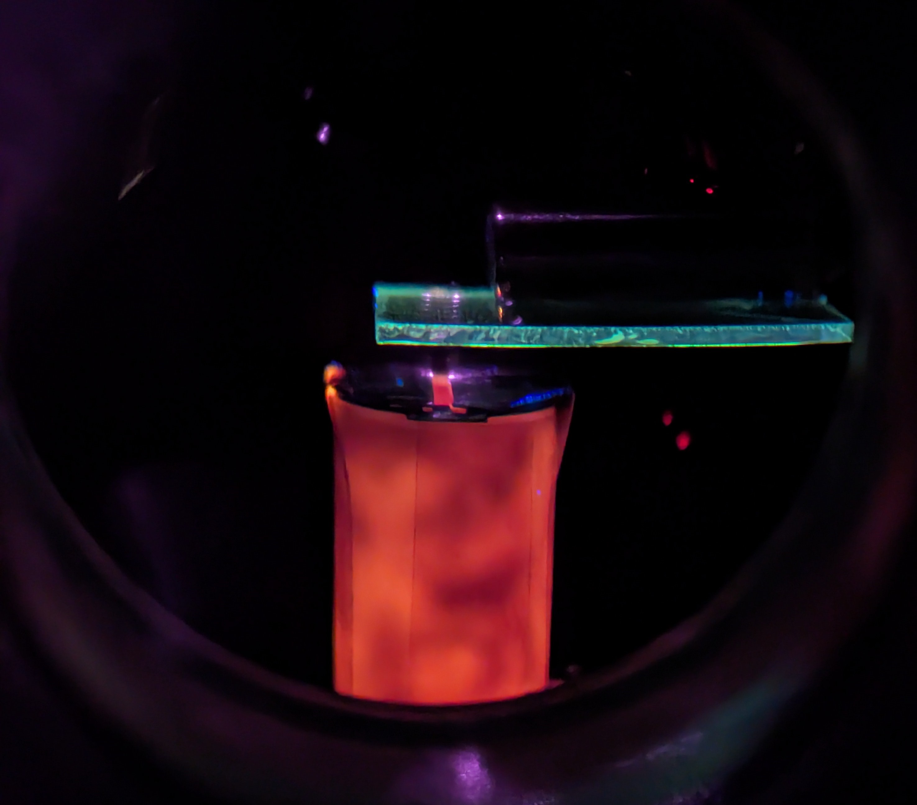
Quantum laser technology developed in our group has provided an unexpected solution to the isotope problem. Drawing on physics work manipulating individual atoms with laser trapping, we developed a way of controlling atoms to separate isotopes with high efficiency and extraordinary purity (over 99.95%). This method, termed Magnetically Activated and Guided Isotope Separation (MAGIS), can produce highly enriched isotopes of numerous elements at relatively low cost and high reliability.
We are exploring the use of fluorescence spectroscopy to detect stable isotopes of iron in blood. Previously developed dry-ashing techniques can be used to extract the iron content of blood samples as iron oxide powder. These iron oxide samples can be vaporized via laser ablation and collimated to produce beams of atomic iron suitable for isotopic spectroscopy. We then resolve the isotope peaks of naturally occurring iron-54, 56, 57, and 58 via fluorescence spectroscopy. Currently, we are working to improve the sample efficiency of our spectroscopic system and thus minimize the required blood volume to produce a diagnostic spectrum, which is an important criterion for applications involving infants and small children.

The work at UT will be further developed in conjunction with the non-profit Pointsman Foundation, where Mark Raizen is Founder and Chairman of the Board, www.pointsman.org. The goal is to translate scientific research into clinical practice. After iron, other essential mineral deficiencies such as zinc and calcium can be addressed with these techniques, to ensure the proper development of at-risk children. Possible future projects also include ultra-sensitive detection of toxic heavy metals, such as lead, cadmium, arsenic, and mercury, which can get into the water or food system and cause serious health problems, and determining exposure is of particular concern in children.
Quantum-Limited Acoustic Imaging
Fundamental physics and medical imaging have long had a close relationship, beginning with the discovery of X-rays in 1895 by Wilhelm Conrad Roentgen. Since then, medical imaging has evolved to include ultrasound, CT scans, MRI, and others- the inventions of which all rely on advances in physics. Improvements in medical imaging over the last 50 years have significantly improved the ability of modern medical professionals to non-invasively detect and diagnose a wide range of ailments.
Like the physicists who walked before us, the Raizen lab proposes an investigation into a novel method of imaging: using the precision and sensitivity of acousto-optic sensing to noninvasively measure vibrations created by energy and heat variations in the body. This idea leverages our recent findings pertaining to the use of a Sagnac interferometer for acoustic detection, as demonstrated by Hillberry et al.
In the proposed method, the position of a laser beam reflected at a glancing angle from a target on a body is used to detect miniscule vibrations on the body's surface. By triangulating multiple targets on the surface, the origin of these vibrations can be located and the intensity measured. Measuring these vibrations provides information on local heating in the body, per the thermoacoustic effect, allowing in situ measurements of the location and magnitude of energy deposited during medical treatments such as Stereotactic Radiosurgery and High Intensity Focused Ultrasound.
Currently, our group is working to determine the maximal sensitivity that could be achieved with this method and comparing it to concurrently developing technologies, such as capacitive micromachined ultrasonic transducers (CMUTs).
Fundamental Physics I: Short-Time Brownian Motion
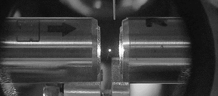
Since 2010, we have built new experiments to trap micron-sized glass beads in liquid, air, and vacuum. We have also built a novel detection system to monitor the real-time position of a trapped bead on very short time and length scales.
One motivation for this work was a long-standing prediction in physics: In 1907, Albert Einstein published a paper where he considered the instantaneous velocity of a Brownian particle and showed that it could be used to test the equipartition theorem, one of the basic tenets of statistical mechanics. However, Einstein concluded that it would be impossible to measure instantaneous velocity in practice due to the very rapid randomization of the motion.
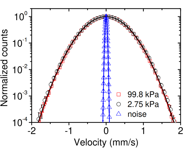
We have measured the instantaneous velocity of a Brownian particle, over 100 years since Einstein's prediction. Our velocity data was used to verify the Maxwell-Boltzmann velocity distribution and the equipartition theorem for a Brownian particle. We have conducted similar measurements in liquid and resolved the instantaneous velocity. The physics in the case of liquid is much richer than air due to viscous fluid effects like memory and added mass.
We have recently used the energy equipartition theorem to "weigh" a microscopic particle in air at the pico-gram level by fitting the mass to the observed Maxwell-Boltzmann distribution. Mass metrology at these levels has applications to single-particle aerosol science including cloud seeding and heterogeneous nucleation of super-cooled liquids, two phenomena that are, at present, poorly understood, but of general interest for climate science.
Also in air, we have demonstrated that optically trapped microbeads are sensitive high bandwidth acoustic transducers. Unlike traditional microphones, which measure acoustic pressure, the micro-bead is sensitive to acoustic velocity. Our micro-bead sensor has superior sensitivity to any commercially available device capable of directly measuring acoustic velocities. Because of the miniscule inertia of these micro-beads, the acoustic sensitivity at high frequencies is also superior to that of commercially available microphones, allowing our sensor to resolve the short rise times of acoustic impulses. Applications range from pinpointing the stopping point of protons in the body during proton therapy for cancer, to detection of acoustic events inside bubble chambers. The latter are being used to search for dark matter and cosmogenic neutrinos.
For Brownian motion in liquids, we are now developing a pulsed laser detection method that we believe will break into the experimentally new regime of compressible-flow Brownian motion. The compressible regime is characterized by time scales shorter than the speed of sound crossing the bead diameter. At these scales, added-mass effects break down and the bead's bare mass enters the equipartition theorem. Therefore, this system will enable the first precise test of the equipartition theorem for a colloidal particle in a classical liquid. We plan to use this system to study thermodynamics and statistical mechanics of small systems out of equilibrium and the emergence of the "arrow of time."
Fundamental Physics II: New Tests of Quantum Mechanics
The Schrödinger equation in quantum mechanics is time-symmetric, while classical dissipative systems have an "arrow of time." The decay of an unstable quantum system is described using a density matrix, and alternatively by quantum trajectories where state collapse is randomly inserted. This framework is a working approach that is confirmed by all experiments to date, but there is a general agreement that the current quantum theory is incomplete since it does not include interactions with the environment in a fundamental way. The key question is whether there are any new experimental tests that could violate the current quantum theory, requiring its revision. This tension has led to recent theoretical models that extend quantum mechanics and naturally incorporate interaction with the environment. We proposed that an atomic clock of a radioisotope could exhibit an "aging effect" where the clock frequency varies over several half-lives as it becomes entangled with the environment.
Specifically, we will search for new effects beyond standard quantum mechanics by measuring the ground state hyperfine splitting of trapped ions, which is proportional to the nuclear magnetic moment. Towards this effort, we will separate a stable isotope of strontium, Sr-88, and then transmute it with a nuclear reactor to produce the radioisotope Sr-89 by neutron capture. This radioisotope has a half-life of around 50.5 days, and decays to Y-89. The measurement time for trapped ion microwave clocks to reach their maximum accuracy is 1-2 days, allowing the precise tracking of the hyperfine splitting over several half-lives.
We will also test whether stable isotopes that were created during nucleosynthesis in the early universe are identical to the same stable isotopes that are newly formed. To test this hypothesis, we will enrich stable Sr-86, transmute it to stable Sr-87 with a nuclear reactor, and separate the newly formed isotope from the target isotope. Two linear ion traps will be loaded with "old" and "new" Sr-87 ions, and we will perform parallel measurements of hyperfine splitting to search for any difference.